


 Foundational characteristics of cancer include proliferation, angiogenesis, migration, evasion of apoptosis, and cellular immortality. Find key markers for these cellular processes and antibodies to detect them.
 The SUMOplot™ Analysis Program predicts and scores sumoylation sites in your protein. SUMOylation is a post-translational modification involved in various cellular processes, such as nuclear-cytosolic transport, transcriptional regulation, apoptosis, protein stability, response to stress, and progression through the cell cycle.
 The Autophagy Receptor Motif Plotter predicts and scores autophagy receptor binding sites in your protein. Identifying proteins connected to this pathway is critical to understanding the role of autophagy in physiological as well as pathological processes such as development, differentiation, neurodegenerative diseases, stress, infection, and cancer.
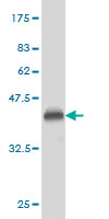
PLD2 Antibody (monoclonal) (M01)
Mouse monoclonal antibody raised against a partial recombinant PLD2.
- SPECIFICATION
- CITATIONS: 3
- PROTOCOLS
- BACKGROUND

Application
| WB, E |
|---|---|
| Primary Accession | O14939 |
| Other Accession | BC015033 |
| Reactivity | Human, Rat |
| Host | mouse |
| Clonality | Monoclonal |
| Isotype | IgG2a Kappa |
| Clone Names | 1C5 |
| Calculated MW | 105987 Da |
| Gene ID | 5338 |
|---|---|
| Other Names | Phospholipase D2, PLD 2, hPLD2, Choline phosphatase 2, PLD1C, Phosphatidylcholine-hydrolyzing phospholipase D2, PLD2 |
| Target/Specificity | PLD2 (AAH15033, 834 a.a. ~ 933 a.a) partial recombinant protein with GST tag. MW of the GST tag alone is 26 KDa. |
| Dilution | WB~~1:500~1000 E~~N/A |
| Format | Clear, colorless solution in phosphate buffered saline, pH 7.2 . |
| Storage | Store at -20°C or lower. Aliquot to avoid repeated freezing and thawing. |
| Precautions | PLD2 Antibody (monoclonal) (M01) is for research use only and not for use in diagnostic or therapeutic procedures. |

Provided below are standard protocols that you may find useful for product applications.
Background
Phosphatidylcholine (PC)-specific phospholipases D (PLDs; EC 3.1.4.4) catalyze the hydrolysis of PC to produce phosphatidic acid and choline. Activation of PC-specific PLDs occurs as a consequence of agonist stimulation of both tyrosine kinase and G protein-coupled receptors. PC-specific PLDs have been proposed to function in regulated secretion, cytoskeletal reorganization, transcriptional regulation, and cell cycle control.
References
1.Construction of lentiviral shRNA expression vector targeting phospholipase D2 (PLD2) gene??.Lian XF, Yu CX, He XL, Lin JJ, Chen YZ, Zhu L.African Journal of Biotechnology Vol. 10 (66), pp. 15044-15050, 26 October, 20112.Mechanisms for the activity of heterocyclic cyclohexanone curcumin derivatives in estrogen receptor negative human breast cancer cell lines.Somers-Edgar TJ, Taurin S, Larsen L, Chandramouli A, Nelson MA, Rosengren RJ.Invest New Drugs. 2009 Oct 9. [Epub ahead of print]
If you have used an Abcepta product and would like to share how it has performed, please click on the "Submit Review" button and provide the requested information. Our staff will examine and post your review and contact you if needed.
If you have any additional inquiries please email technical services at tech@abcepta.com.
Ordering Information
Other Products
Shipping Information
